Plant Leaf Diseases: Spots, Yellowing, Mildew, and Damage Clues
Learn which photo clues actually help diagnose plant leaf diseases, which signs are misleading, and the safe next steps to investigate before acting.
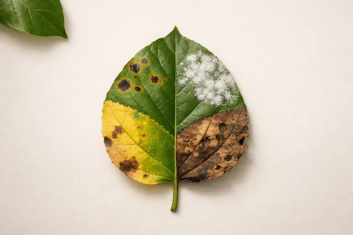
At a glance: what leaf clues reveal about plant leaf diseases
Plant leaf diseases often leave distinctive visible clues, but a single photo rarely proves a specific pathogen. Photos are best at showing whether a leaf has spots, patterns of yellowing, powdery growth, or physical damage — and those patterns help you narrow the set of likely causes.
Use photos to confirm pattern-level evidence: whether lesions are circular or irregular, whether yellowing follows veins or appears across whole leaves, and whether powdery coatings wipe off. Those observations tell you which category of disease or stress is most plausible, for example fungal leaf spot, bacterial blight, nutrient deficiency, or insect feeding.
Treat any single-photo diagnosis as tentative. The strongest outcome from one image is a prioritized list of possibilities and a short, safe next-step checklist (look at other plants, check stems and soil, note timing and weather). That approach reduces wasted treatments and avoids the risks of applying fungicides, fertilizers, or pesticides based on uncertain evidence.
If your goal is action, prioritize containment and low-risk responses: isolate an affected plant if feasible, remove badly affected leaves, improve airflow, and document changes over a few days. Use the clues below to choose which of those steps to try first and when to escalate to lab testing or professional advice.
- Photos reliably show patterns (spots, margins, powder, bite marks) but not the exact pathogen.
- Look for repeatable patterns across multiple leaves and plants before treating.
- Record context: plant species, recent weather, watering schedule, and nearby pests.
- Use low-risk, reversible actions (prune, clean, increase airflow) while you collect more evidence.
Most reliable visual clues for plant leaf diseases
Some clues hold up well even in imperfect phone photos. Shape and distribution of lesions — for example discrete round spots with concentric rings, angular lesions that stop at veins, or necrotic patches along the leaf edge — are stronger evidence than color alone. Those geometric patterns map to common causes: fungal leaf spots often form round or target-like lesions, bacterial infections can create water-soaked or angular spots, and abiotic chemical burns often affect margins and tips first.
Surface texture is another durable clue. Powdery or downy mildew leaves a distinct powdery or fuzzy white/gray coating that usually rubs off. Scab-like crusts, sticky bacterial exudate, or raised pustules suggest specific pathogen groups. Take a close, in-focus shot of the affected surface and try to include a fingertip or coin for scale so you can judge texture.
Vein-associated patterns are informative. Chlorosis that radiates from the veins, or interveinal yellowing with green veins, often indicates nutrient imbalance or systemic infection. Conversely, lesions that are restricted by veins and appear angular often point to bacterial or some fungal leaf spots because veins act as physical barriers to fungal spread.
Pattern across the plant is essential. When many leaves across multiple stems show similar spots at the same stage, an infectious spread is more likely than a localized injury or isolated nutrient deficiency. Pair leaf photos with shots of the whole plant and neighboring plants to confirm whether the problem is plant-specific or environmental.
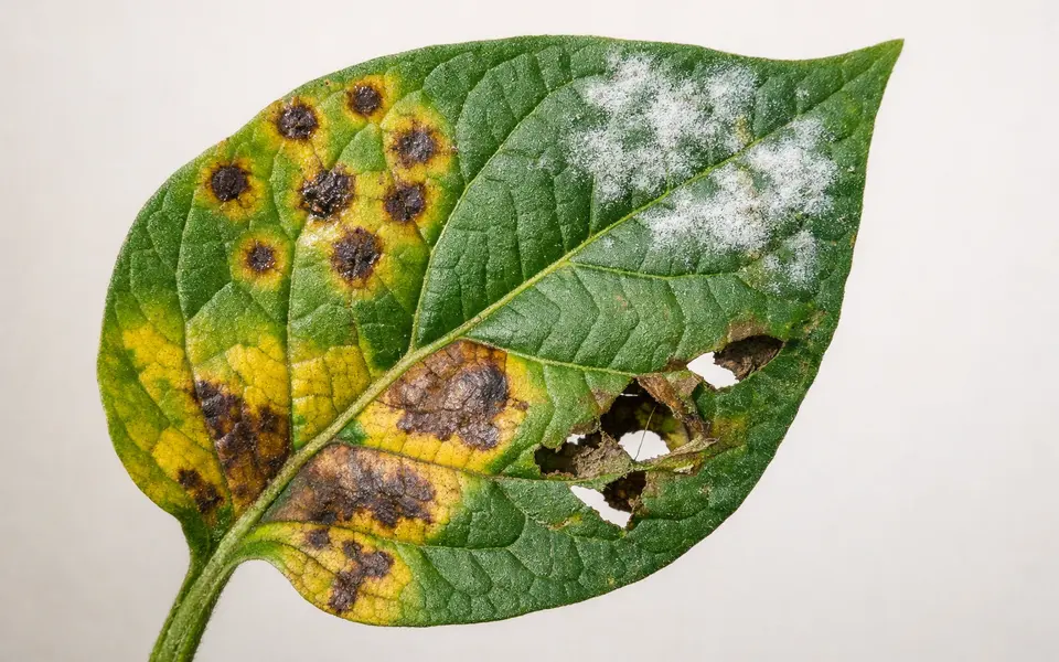
- Lesion geometry: round rings, angular spots, margins, or streaks that suggest pathogen type.
- Surface texture: powdery, fuzzy, sticky, or crusty details that identify mildew, bacteria, or scale.
- Vein relationship: interveinal yellowing vs. vein-limited lesions helps separate nutrient vs. infectious causes.
- Distribution pattern: single leaf, many leaves, or multiple plants indicates local injury vs. contagion.
- Scale and timing: include an object for size and note whether symptoms appeared gradually or suddenly.
Weak or misleading signs for plant leaf diseases
Color alone is a weak signal. Lighting, camera white balance, and leaf age change how greens and yellows appear in photos. Relying on a photo’s color to decide between nutrient deficiency and disease is risky; instead, combine color with texture and pattern clues. For example, both magnesium deficiency and certain viral infections can cause yellowing, but viruses more often produce banding or mosaic patterns rather than uniformly dull leaves.
Single-angle photos and closeups that miss context commonly mislead. A close macro of a brown spot can look like a fungal lesion but may be a healed insect wound or physical scorch. Always photograph the leaf from multiple angles: top surface, underside, the petiole, and the whole plant. Undersides often show insect eggs, scale insects, or spore-producing structures that aren’t visible on top.
Shiny glare and backlighting can hide texture. Powdery mildew may disappear in a glare-heavy shot, while water droplets can mimic fungal sporulation. If possible, take photos in diffused daylight, avoid flash, and gently blot water before photographing. If glare is unavoidable, capture a second image with a different angle or include a tactile note (does the coating brush off?).
Single-plant symptoms without environmental context can point in the wrong direction. Soil pH, recent fertilizer application, herbicide drift, and root damage all produce leaf-level symptoms but require different interventions than contagious diseases. If a problem emerged right after a new fertilizer or nearby spraying, consider abiotic causes first and collect that history before assuming an infectious disease.
- Don’t trust hue differences alone—camera and light affect color.
- Avoid diagnosing from a single closeup; get top, bottom, and whole-plant views.
- Watch for glare, shadows, and reflections that hide surface texture.
- Consider recent care actions (fertilizer, sprays, transplanting) as likely noninfectious causes.
- Be skeptical of symptom names from web image matches without follow-up clues.
How to compare multiple clues to narrow plant leaf diseases
Build a short comparison checklist: pattern geometry, surface texture, vein relationship, distribution, and recent context. Take photos that cover each checklist item and then score how many clues point to each likely cause. That method turns a vague visual match into a reasoned shortlist: for example, round spots + concentric rings + many affected leaves suggests fungal leaf spot; angular, vein-limited lesions + water-soaked look suggests bacterial infection.
Work through elimination. If powdery coating is present and rubs off, deprioritize nutrient deficiency. If lesions are isolated to leaf tips and margins with uniform yellowing beginning at edges, give precedence to drought, salt, or herbicide damage. Keep a running list of matches and mismatches instead of forcing a single answer—most practical decisions require knowing which causes are most probable and which are unlikely.
Use the plant and season as tiebreakers. Some diseases are seasonal (e.g., downy mildew in cool, wet springs), while others flourish in heat and humidity. Certain hosts are vulnerable to predictable pathogens. A host plant that’s commonly affected by powdery mildew narrows the candidate list more than looking at a leaf shape alone. When possible, annotate photos with date, recent weather, and plant variety to make comparisons more reliable.
If you want a second reference point, compare your findings to a trusted checklist. Our Plant Identification Cheat Sheet (https://leafidentification.app/blog/plant-identification-cheat-sheet) complements this article by listing lookalikes and verification steps for visual matches. Use that resource to test whether the clues you’ve collected fit a documented pattern or whether they’re ambiguous enough to require lab testing.
- Collect photos for each checklist item: texture, geometry, vein relation, distribution, and context.
- Score matches: three or more consistent clues increases confidence in a likely category.
- Eliminate options that conflict with strong clues (e.g., powder that wipes off rules out many nutrient problems).
- Use season and host susceptibility as practical tiebreakers.
- Refer to a trusted checklist to spot common lookalikes before choosing treatments.
Using Leafzy after you check plant leaf disease clues
Once you’ve collected the core clues, use Leaf Identification: Leafzy as a structured next step. The app is most useful for turning your photo-based checklist into candidate matches and showing side-by-side examples to compare texture and distribution. Treat app results as prioritized suggestions to guide further observation, not as final proof of a specific pathogen.
Open the app with a set of clear photos: an in-focus closeup of the lesion surface, a shot of the leaf underside, and a whole-plant view that shows how widespread symptoms are. Add notes within the app or your phone’s camera app describing recent watering, sprays, temperature, and whether other plants are affected. Those context notes improve how you interpret suggested matches.
If Leafzy suggests several possibilities, refer back to your checklist and try quick verification steps that are safe and reversible: gently wipe suspected powdery growth to see if it rubs off, check for tiny insects or eggs under the leaf, and observe whether symptoms spread to adjacent plants over a few days. If matched possibilities require treatment, prefer targeted, low-impact measures first—hand removal, pruning, improved airflow, and adjusting irrigation—until a confident diagnosis is made.
When results remain uncertain or the plant is valuable, consider lab testing or professional diagnosis. Keep a record of the app suggestions and the clues that led you there; professional labs and advisers can use that history to speed up verification. Remember that the app helps you prioritize what to test and which low-risk actions to take first.
- Take 3 photos for the app: lesion closeup, underside, and whole-plant context.
- Include notes about recent care, weather, and affected neighbors before scanning.
- Use app matches as candidates—then run quick, reversible checks (wipe, inspect underside, isolate plant).
- Start with low-impact responses; escalate to pesticides or lab tests only if evidence points there.
- Document app suggestions and follow-up observations for later reference or professional review.
Related guides
Next step: check clues, then scan with Leaf Identification: Leafzy
After you collect clear photos (lesion closeup, underside, whole-plant) and note recent care and weather, use Leaf Identification: Leafzy to turn your checklist into prioritized candidate matches. Treat app suggestions as research notes: run quick verification checks (wipe off powdery growth, inspect undersides, isolate affected plants) and prefer low-impact responses while you gather more evidence. If the app flags multiple possibilities or symptoms worsen, save the app results and consider professional advice or lab testing before using stronger treatments.

Frequently asked questions
Can I identify a specific pathogen from a single leaf photo?
A single leaf photo rarely proves a specific pathogen. Photos are excellent for showing patterns—spots, powder, vein-related yellowing—but multiple clues and context are needed to narrow a diagnosis. Treat any single-photo match as a prioritized possibility and follow up with more photos, environmental notes, and simple verification checks before applying targeted treatments.
What quick checks can confirm whether a coating is powdery mildew?
Powdery mildew typically forms a dry, powdery or dusty white to gray coating that often wipes away with a fingertip or soft cloth. Take a close shot and then gently brush the surface; if the powder smudges or rubs off and the leaf underneath looks intact, mildew is likely. Also check the underside of leaves and nearby plants for similar coatings.
How do I tell if yellowing is a disease or a nutrient problem?
Look at the pattern: interveinal yellowing with green veins often points to nutrient deficiencies (e.g., magnesium or iron), while irregular yellow patches or yellowing accompanied by discrete lesions suggests disease. Consider timing, recent fertilization, and whether the problem affects many plants or just one. If in doubt, correctable nutrient remedies (minor foliar feed or soil adjustment) are low-risk tests compared with broad chemical treatments.
When should I send a sample for lab testing or call a professional?
Consider lab testing or professional help when the plant is high-value, symptoms are spreading rapidly despite reasonable low-impact measures, or you need a definitive identification before using stronger treatments. If symptoms persist, worsen, or involve woody stems and roots, collect representative samples, document the progression, and share your app notes and photos with the lab or advisor to accelerate diagnosis.